Diagnostic Tests
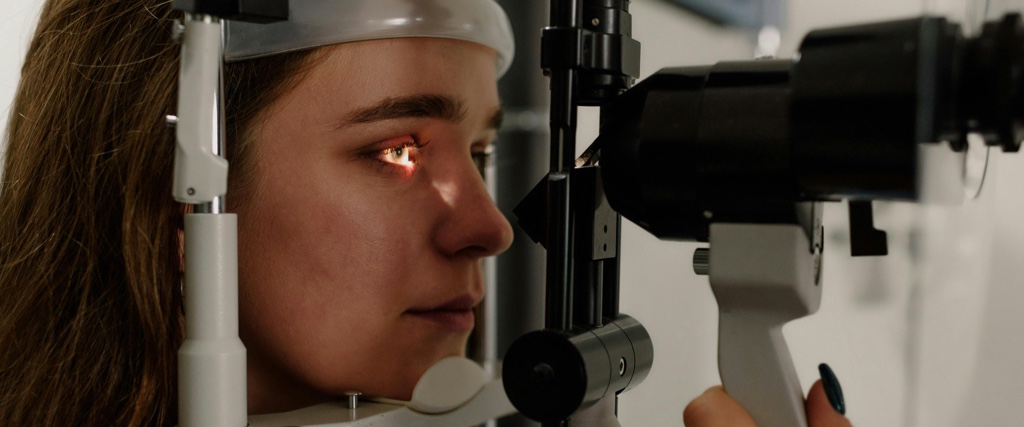
External Photography
External photography allows us to document and demonstrate changes in the superficial structures of the eye and eyelids. The camera is mounted to a slit lamp and has a monitor that allows us to show the patient and family the objects of interest, thus improving patient comprehension and alleviating anxiety.
Gonioscopy
Gonioscopy is an examination used to evaluate the internal drainage system of the eye, or the trabecular meshwork, and its anatomical relation to the adjacent iris. A special contact lens mirror is used to visualize these structures, which are hidden from direct observation. Gonioscopy is a painless examination used to determine whether internal drain or drainage angle is either open or closed. Depending on your age and whether you are at high risk for developing glaucoma, gonioscopy may be a routine part of your evaluation.
Lenstar
Accuracy in IOL power selection optimizes your vision after cataract surgery and helps reduce dependence upon glasses.Lenstar provides laser interferometric biometry of the entire eye, precise keratometry, astigmatism and axis measurement, white to white and pupillometry simultaneously in a single measurement.
OCT
Cirrus HD OCT uses infrared light to obtain a detailed and precise image of the retina and optic nerve. Analogous to a CT scan, the OCT allows detection of microscopic changes before they become visible to an examiner’s eye. This technology aids in the diagnosis of retinal diseases, as well as determining whether glaucoma is adequately controlled.
Orbscan Corneal Topography
An Orbscan is a diagnostic test that measures the curvature and thickness of your cornea (the clear window of your eye). This “no touch” procedure quickly completes 10,000 measurements and determines subtle irregularities in the shape of your cornea that cannot be determined by standard examination techniques. The Orbscan is critical in determining your suitability for laser refractive surgeryRetinal Photography
A special camera that digitally records images of the retina, the retinal blood vessels, and the optic nerve. This allows your doctors to follow and treat glaucoma, diabetic retinopathy, and macular degeneration with great accuracy.Specular Microscopy
Specular Microscopy measures the number, shape, and size of the cells on the inside surface of your cornea. These cells maintain corneal clarity and good vision. Trauma and some inherited conditions may cause a loss of these cells and an accurate assessment of their health is crucial.Visual Fields
The Humphrey Visual Field Analyzer measures hundreds of spots in your side (peripheral) vision, usually within 4 minutes per eye, detecting and monitoring glaucoma, strokes and tumors. This “smart test” also determines your ability to provide consistent responses, filtering out misleading data.OPD Scan III (Nidek)
The OPD Scan III identifies and measures irregularity in the cornea and in the physiologic lens that allows us to decide the most appropriate surgical plan to restore optimum vision. It tells us whether you are best suited for cornea refractive surgery or lens-based refractive surgery by evaluating your total visual system.